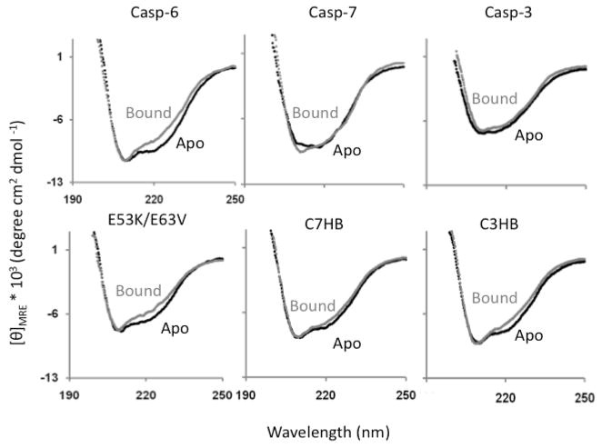
FIGURE 2.
CD spectra of apo and ligand-bound wild-type caspase-6 reflect the change in structure of the 60’s and 130’s helix upon ligand binding. In contrast the CD spectra of caspase-7 and -3 do not reflect a change in helical content. The network-disrupting variant (E53K/E63V) shows a similar change in helical content upon ligand binding to wild-type caspase-6. The helix breaking variants C7HB and C3HB show a much decreased difference in CD spectra between apo and ligand bound, suggesting that the apo state is less helical in solution than wild-type caspase-6.

