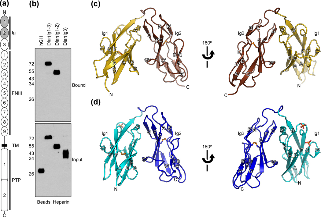
Fig. 1.
Crystal structures of the Ig1–Ig2 domains of Dlar and mouse LAR. (a) Schematic representation of type IIa RPTPs. Ig: immunoglobulin-like domains, FNIII: fibronectin type III domains, TM: transmembrane region, PTP: protein tyrosine phosphatase domains. The Ig domains that were crystallized are shaded. (b) Identification of a heparin-binding region within the Ig domains of Dlar. Fragments of Dlar were fused to human growth hormone (hGH) and expressed transiently in HEK293 cells. Conditioned media were incubated with heparin-sepharose. Bound fusion proteins were visualized by immunoblotting against hGH. (c) Ribbon diagram of Dlar(Ig1-2). The letters N and C indicate the N- and C-termini, respectively. Each β-strand is labeled. Disulfide bonds are shown as orange ball-and-stick models. The first and second Ig domains are colored gold and brown, respectively. (d) Ribbon diagram of mouse LAR(Ig1-2). The first and second Ig domains are colored cyan and blue, respectively. Two bound sulfate ions are shown in ball-and-stick representation. Oxygen and sulfur atoms are colored red and orange, respectively. Structural images were prepared with PYMOL (www.pymol.org).

