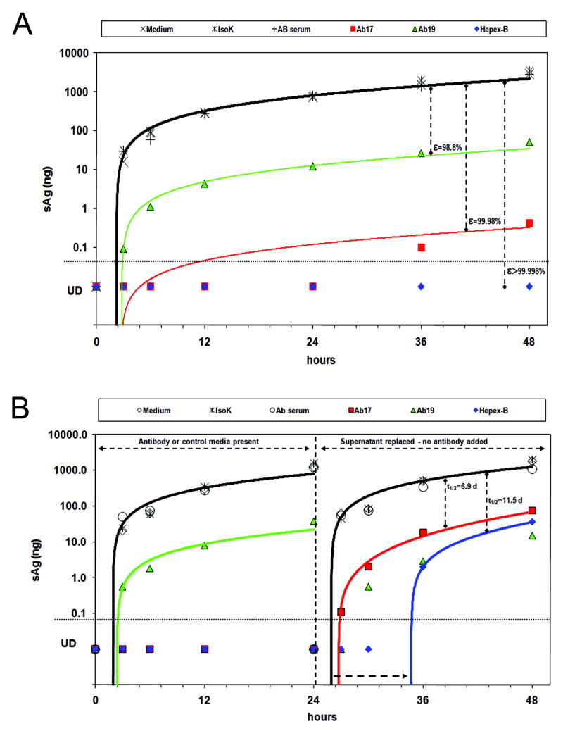
Figure 5. Supernatant HBsAg kinetics in-vitro shows blocking of particle release by PLC/PRF/5 cells incubated with Hepex-B™.
A) Significantly lower HBsAg levels were observed when cells were incubated for 48 hours with HBV-Ab 17 (red squares), HBV-Ab 19 (green triangles) or Hepex-B™ (blue diamonds) as compared to medium (empty diamonds), isotype-control (stars) or human AB serum (empty circles). Fitting with the mathematical model (solid lines) indicates blocking of particle release. B) Cells were incubated with mAbs for 24 hrs, after which the supernatants were replaced with medium only. Blocking release continued in the absence of anti-HBs in the supernatant, with a gradual decline of its effectiveness.

