
Figure 4.
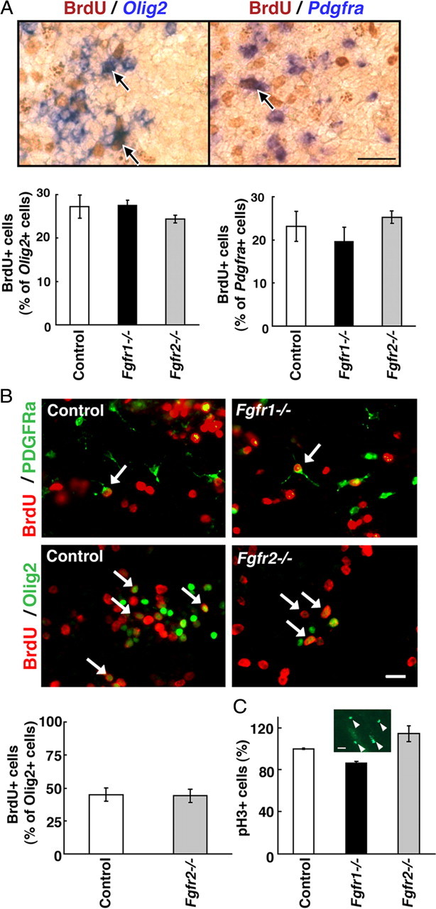
Loss of Fgfr1 or Fgfr2 does not affect proliferation of OL progenitors. A, Mothers were injected intraperitoneally with BrdU (100 mg/kg body weight) 1 h before harvesting the embryos. To identify proliferating cells, coronal sections of E12.5 forebrain from Fgfr1−/− or Fgfr2−/− mutants and littermate control were double labeled by combining immunohistochemistry for BrdU (brown) and in situ hybridization for Olig2 or Pdgfra mRNA expression (purple). Total numbers of BrdU+ cells double labeled for Pdgfra or Olig2 mRNA (arrows) were counted in the entire section as in Figure 1 and expressed as percentage of Olig2- or Pdgfra-positive cells. No significant differences were observed in the numbers of proliferating OLPs between controls and either of the mutants. Error bars represent SEM (N = 3–4). B, Dissociated cell cultures initiated from E12.5 forebrains of control, Fgfr1−/−, or Fgfr2−/− embryos were treated with BrdU (50 μm) for 3 h and cells were double immunolabeled with anti-BrdU and either anti-Olig2 (3 DIV) or anti-PDGFRa (6 DIV). No differences were observed in the numbers of Olig2+ cells incorporating BrdU in control and Fgfr2−/− (quantified) or Fgfr1−/− mutant cultures. The arrows show examples of double-labeled cells. Error bar represents SEM for control (N = 3) and means of the difference for Fgfr2−/− (N = 2). C, Coronal sections of E12.5 forebrain from control, Fgfr1−/−, or Fgfr2−/− mice were immunostained with anti-pH3 (a marker for mitotic cells). Total numbers of pH3-positive scattered cells were counted in the entire section as for Figure 1 [representative area with pH3+ cells (arrowheads) is shown in inset], and the numbers were expressed as a percentage of the control. No differences in the numbers of pH3+ cells were observed between the control and mutants. Error bars represent mean of duplicates. Scale bars: A, 50 μm; B, 20 μm; C, 20 μm.