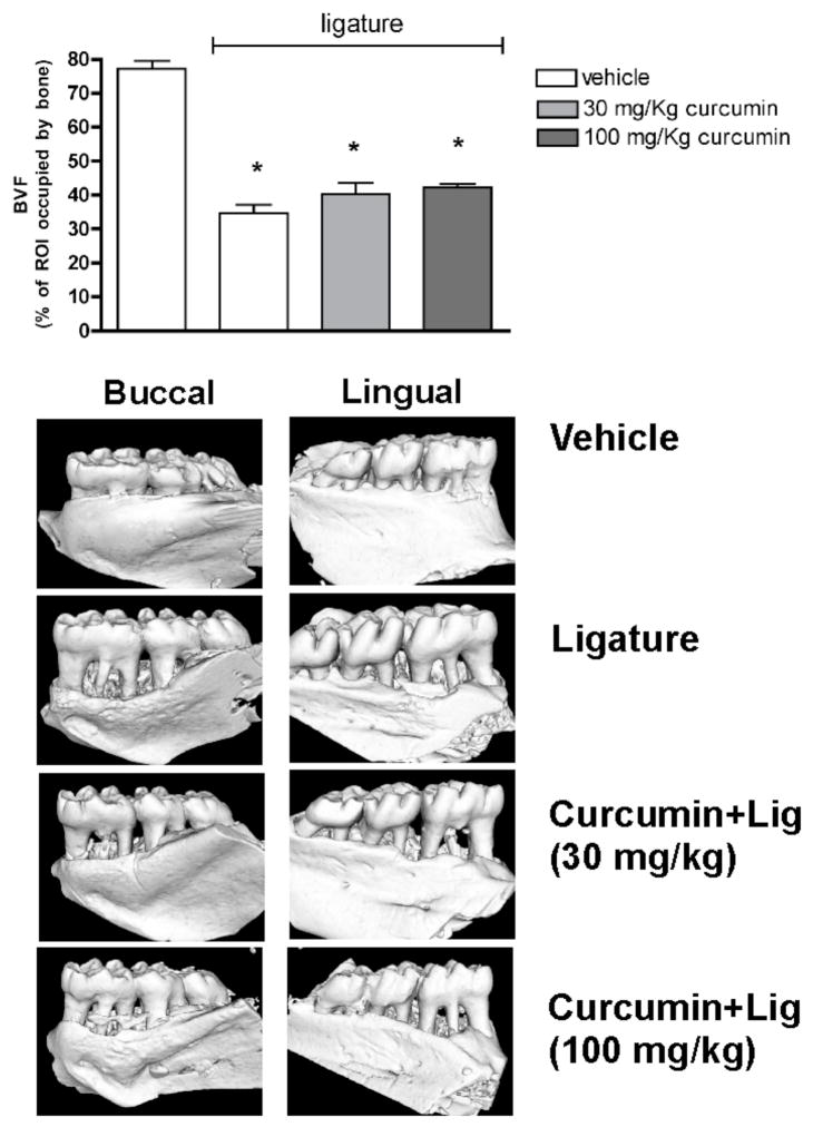
Figure 1.
μCT analysis of mandible specimens obtained from the experimental animals shows that neither dose of intragastrically-administered curcumin prevented alveolar bone loss in the ligature-model of periodontal disease. The tridimensional images are representative of 8 samples in each group (4 animals/group) and depict the significant resorption of alveolar bone on both the buccal and lingual aspects of the first molars. The bar graphs represent the quantification of the area occupied by bone tissue in a standardized region of interest (ROI) defined by anatomic landarks including the first molar and the interproximal area between first and second molars. Bars represent means and vertical lines standard deviations of the percentage of the ROI occupied by bone tissue. *indicates a significant (p<0.05) difference in comparison with negative control specimens from animals treated with vehicle and without ligatures.

