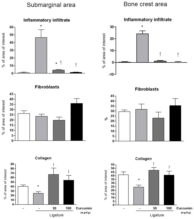
Figure 2.
Stereometry analysis of the histological characteristics of gingival tissues subjected to ligature-induced periodontal disease according to the dose of curcumin (30 or 100 mg/Kg) administered for 15 days intragastrically. Control animals were administered vehicle and negative control animals also were given vehicle but no ligature was placed around the first molars. Two 50 × 50 μM grids were overlayed on histological images captured from H/E stained slides at 200 X magnification, allowing the analysis of two ‘areas of interest’ of 2,500 μm2 each: one was positioned 25 μM below the top of the alveolar bone crest, and another at the base of the junctional epithelium, representing the ‘bone crest’ and ‘submarginal’ areas, respectively. The proportion of collagen, fibroblastic cells and inflammatory cells (distinguished by the morphological characteristics) in the area of interest was determined. A total of three images obtained from equally spaced slides (spanning 900 uM of the buccal-lingual aspect of the molars) were evaluated from each animal. Slides from at least three different animals in each experimental group were used. Both doses of curcumin markedly reduced the inflammatory infiltrate in periodontally-diseased tissues. The lower dose of curcumin (30 mg/Kg) induced a greater increase in the collagen content, whereas the higher dose (100 mg/Kg) was associated with increased proliferation of fibroblastic cells. Bars indicate averages and vertical lines the standard deviations. Student’s t-test was used for pairwise comparison and (*) indicates a significant (p<0.05) difference in comparison with healthy (no ligature) control, whereas (!) indicates significant difference (p<0.05) between curcumin-treated and vehicle-treated in periodontal disease (ligature) sites.

