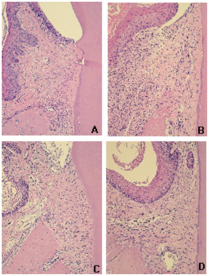
Figure 3.

Histological aspect of the gingival tissues according to the experimental group (control X ligature) and administration of curcumin (vehicle, 30 mg/Kg and 100 mg/Kg). Tissues were harvested 15 days after ligatures were placed and administration of curcumin was started. Semi-serial sections with 5 μM thickness were routinely processed and stained with hematoxylin and eosin. A total of three sections spaced 100 μM were evaluated for each first molar in a minimum of four animals in each experimental group. An epithelial layer of regular thickness, dense connective tissue with reduced number of cellular infiltrate and a smooth bone crest surface characterize the gingival tissues of control (non-ligated) animals (A). Placement of ligatures (B) produced an increase of the thickness of the epithelium layer, an intense cellular infiltrate and irregular bone crest surface with the presence of multinucleated osteoclast-like cells; whereas in animals treated with 30 mg/Kg (C) and 100 mg/Kg (D) of curcumin, the epithelial layer presents normal thickness and reduced cellular infiltrate. All images were obtained at 100 X magnification.
