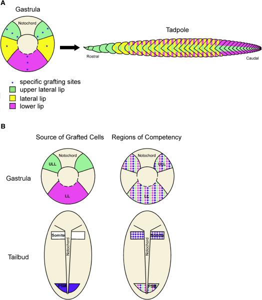
Figure 10. A mesodermal fate map and competence map of gastrula and PSM tailbud cells.
(A) A fate map of the tadpole musculture projected onto the gastrula shows the following: upper lateral lip cells (green) give rise to the anterior-most somites as well as myotome fibers positioned in the central region of somites along the entire axis; cells positioned in the lateral circumblastoporal region (yellow) give rise to myotome fibers found both in the central and dorsoventral regions of somites throughout the trunk region; and lower lip cells (purple) form myotome fibers found in the dorsal and ventral domains of somites located in the trunk and tail regions of the tadpole. (B) A diagram summarizing the competency of gastrula and tailbud PSM cells to give rise to myotome fibers when grafted to different locations. Gastrula cells from the upper lateral (green) and lower (purple) lip regions are competent to give rise to myotome fibers when grafted to any region of the gastrula and to the PSM of tailbud-staged embryos (Fig. 10B; Regions of Competence). Similarly, cells from the PSM of tailbud embryos (blue) are competent to form myotome fibers when transplanted to the gastrula and tailbud PSM. However, PSM cells are also competent to form myotome fibers when transplanted to the anterior end of the paraxial mesoderm whereas gastrula cells are not.

