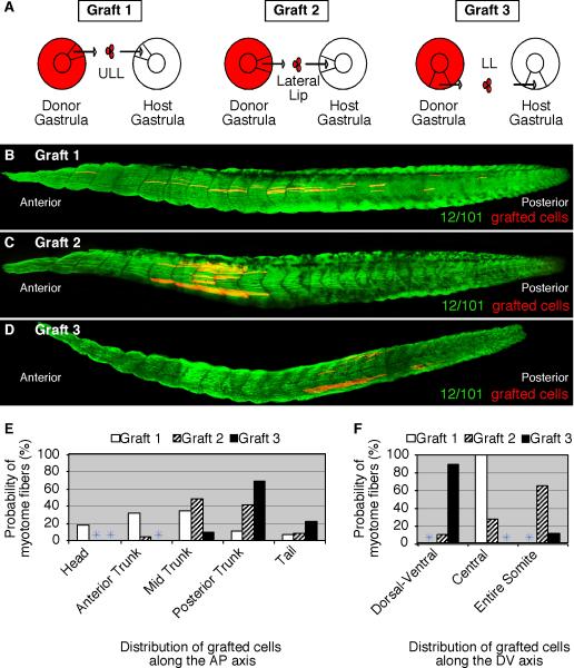
Figure 2. Gastrula cells from different blastopore lip regions form myotome fibers in distinct locations within somites and along the anteroposterior axis.
(A) A diagram depicting homotopic RDA-labeled upper lateral lip-ULL cells (Graft 1), lateral lip cells (Graft 2), and lower lip-LL cells (Graft 3) grafted from stage 11 donor embryos to the same region of unlabeled host embryos. Grafted embryos were allowed to develop to tadpoles (stage 39) and subsequently stained for the myotome-specific marker, 12/101 (green). (B) Grafted ULL cells formed myotome fibers located in the central domain of somites along the entire anteroposterior axis. (C) Grafted lateral lip cells formed myotome fibers found throughout somites position in the trunk whereas grafted lower lip-LL cells formed myotome fibers predominantly in the dorsal and ventral regions of somites in the posterior trunk and tail (D). The probability of ULL (Graft 1), lateral (Graft 2), and LL (Graft 3) cells that give rise to myotome fibers at specific regions along the anteroposterior (E) and dorsoventral (F) axes are shown.

