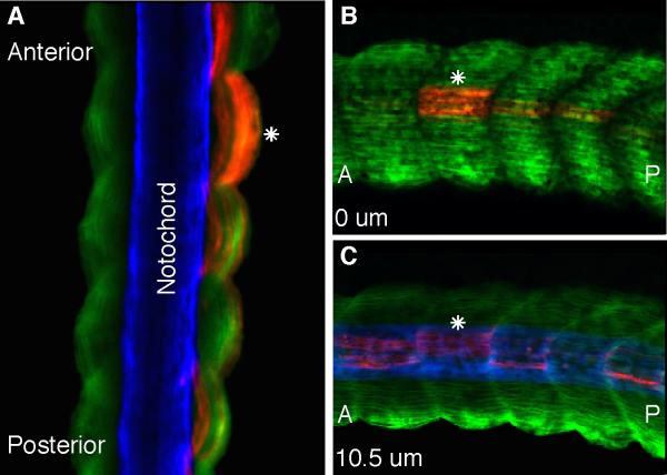
Figure 3. Upper lateral lip cells form myotome fibers in both the medial and lateral aspects of somites.
(A) A dorsal confocal scan of a stage 39 homotopically grafted embryo containing RDA-labeled upper lateral lip cells (red) that have differentiated into myotome fibers as shown by the myotome-specific antibody 12/101 (green). A white asterisk shows labeled myotome fibers are positioned medially, adjacent to the notochord as shown by the Tor-70 antibody (blue) as well as at the lateral edge of the paraxial mesoderm. Lateral to medial confocal scans of the same embryo shows labeled myotome fibers at the lateral (B) and medial (C) edges of somites. (A) anterior; (P) posterior.

