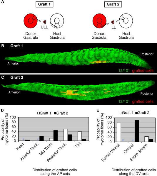
Figure 5. Gastrula cells form myotome fibers according to their new host location.
(A) A diagram depicting heterotopic grafts in which RDA-labeled upper lateral lip-ULL cells are grafted to the lower lip region (Graft 1) and RDA-labeled lower-lip-LL cells are grafted to the upper lateral lip region (Graft 2) of stage 11 host embryos. Grafted embryos were allowed to develop to tadpoles (stage 39) and then subsequently stained for the myotome-specific marker, 12/101 (green). (B) ULL-labeled gastrula cells transplanted to the lower lip region form myotome fibers in the ventral region of somites positioned towards the posterior axis of the tadpole. (C) LL-labeled gastrula cells transplanted to the upper lateral lip region form myotome fibers in the central domain of somites positioned along the axis. The probability of ULL (Graft 1) and LL (Graft 2) cells that give rise to myotome fibers at specific regions along the anteroposterior (D) and dorsoventral (E) axes are shown.

