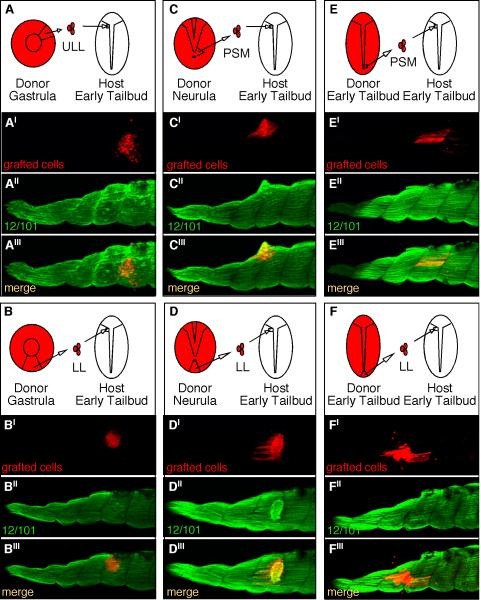
Figure 7. Mesodermal cells gradually become competent to form myotome fibers when grafted to the anterior paraxial mesoderm of early tailbud-staged embryos.
RDA-labeled cells grafted from the gastrula upper lateral lip-ULL (A), gastrula lower lip-LL (B), neurula PSM of a stage 15 embryo (C), neurula tailbud lower-lip of a stage 15 embryo (D), tailbud PSM of a stage 19 embryo (E), and the lower lip of a stage 19 embryo (F) were all grafted to the anterior paraxial mesoderm of stage 19 tailbud embryos where somites have begun to form. Grafted embryos were cultured to stage 39 and then stained with the myotome marker, 12/101 (A″–F″) to determine whether the RDA-labeled cells (A′–F′) had adopted a myotome fate and morphology as shown by the merged images (A″′–F″′).

