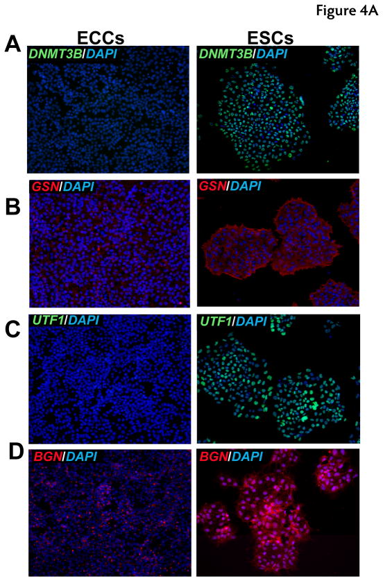
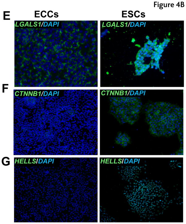
Fig. 4. Immunocytochemical analysis of proteins expressed at high levels in ESCs.
Indirect immunofluorescence labeling of different cell types was carried out using Alexa Fluor 594 or Alexa Fluor 488 conjugated secondary antibodies. DAPI (blue) was used to stain nuclei. Panels A to H show proteins found to be expressed at higher levels in ESCs. Panel A to G includes immunocytochemical staining for proteins encoded by DNMT3B (blue-green nuclei), DNMT3A (blue-green nuclei), GSN (red cytoplasm), UTF1 (blue-green nuclei), BGN (red secretory), LGALS1 (green cell surface), CTNNB1 (green cell surface) and HELLS (blue-green nuclei), respectively.


