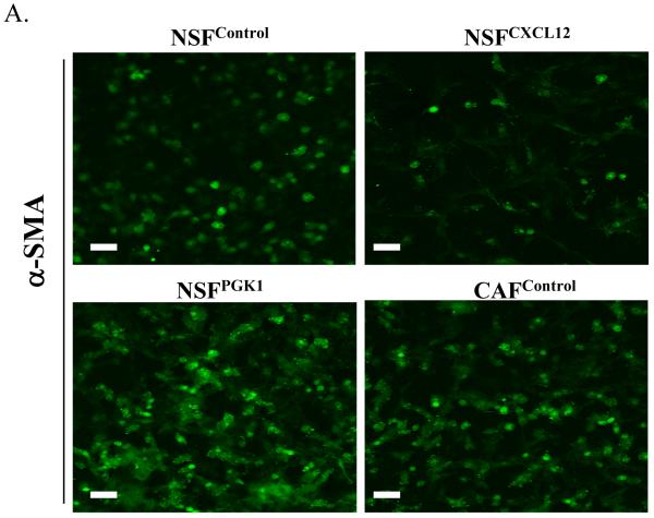
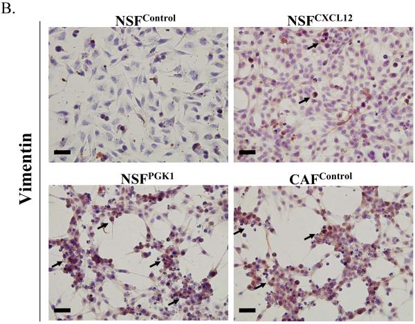
Figure 4. Expression of CXCL12 or PGK1 alters the phenotype of NSFs.
(A) Immunofluorescence staining for α-SMA in NSFCXCL12, NSFPGK1, NSFControl, and CAFcontrol cells Expression of α-SMA (green) is higher in NSFPGK1 and CAFcontrol cells than in NSFControl cells. Original magnification 40×, where the bars represents 50 μM.
(B) Vimentin staining of NSFCXCL12, NSFPGK1, NSFControl, and CAFcontrol cells. Representative immunohistochemical staining for vimentin (brown, nuclear) is presented. Original magnification 40×, where the black bars represents 50 μM.


