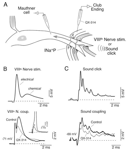
Figure 7. A mechanism of lateral excitation contributes to afferent synchronization.
A, Dendritic synaptic potentials (Mauthner cell) evoked by suprathreshold electrical or natural stimulation of VIIIth nerve afferents (darker afferents; VIIIth Nerve stim. and Sound click; upper traces in B and C) can be recorded as coupling potentials, in neighbouring subthreshold terminals (light terminals; lower traces in B and C, respectively). INa+P (persistent, subthreshold, Na+ current. B, Mixed synaptic response (electrical and chemical) produced by extracellular electrical stimulation of the posterior branch of the VIIIth nerve (Upper, VIIIth Nerve stim.) evokes a retrograde coupling potential in a subthreshold terminal (lower, VIIIth Nerve coup.). Inset: amplitude of the VIIIth nerve coupling (asterisk) was adjusted to be at the threshold of the presynaptic afferent (truncated spike: 100 mV). Superimposed traces in the lower panel represent the amplitude of these retrograde responses obtained right after (control) and 5 minutes after the penetration of the terminal with an electrode containing QX-314. C, Sound-evoked synaptic potential (Sound click; 500 μs pulse) can also be recorded as a coupling potential in neighbouring inactive terminals (bottom trace; Sound coupling). Superimposed traces represent the retrograde responses obtained right after (Control) and 5 min. after the penetration of the synaptic terminal with an electrode containing QX-314. Note the reduction in amplitude of both retrograde coupling potentials observed after injection of QX-314 (grey traces). Modified from Curti and Pereda (2004), with permission.

