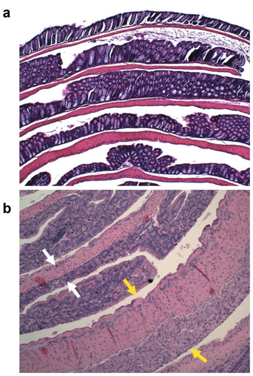
FIG. 4.

Hematoxylin and eosin–stained histologic sections of colons from normal (a) and DSS-treated mice (b) prepared as Swiss rolls. Both images are displayed in the same scale, ×10. In the inflamed colon, white arrows indicate an area with healed epithelium and normal colon wall thickness. The yellow arrows indicate a colonic segment with complete ulceration. The mucosa is replaced by edematous granulation tissue and the colonic wall is markedly thickened.
