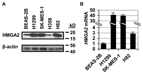
FIGURE 1.
HMGA2 expression is increased in lung cancer cell lines from metastatic disease.
A. Western blot analysis shows increased HMGA2 in the human lung cancer cell lines H1299 (metastatic non-small cell carcinoma), SK-MES-1 (metastatic squamous cell carcinoma), H82 (metastatic small cell carcinoma) compared with the normal human bronchial epithelium cell line BEAS-2B. In H358 cells, derived from non-metastatic, non-small cell bronchioloalveolar carcinoma, HMGA2 protein levels are similar to that of the BEAS-2B immortalized control cells line.
B. Quantitative RT-PCR analysis shows increased HMGA2 mRNA in H1299, SK-MES-1, and H82 metastatic lung cancer cells compared to BEAS-2B cells, consistent with the Western blot analysis (A). Values shown are fold-differences of RNA expression compared to BEAS-2B cells (arbitrarily assigned a value of 1); results represent the mean +/− the standard deviation from two independent experiments performed in triplicate.

