Figure 2.
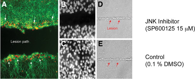
JNK signaling during SE regeneration. JNK signaling is evident at the leading edge of the lesion path in the SEs and necessary for proliferative regeneration. SE cultured on a glass coverslip was lesioned by microbeam laser ablation. A, Phosphorylated c-JUN was detected by a phosphorylation-specific antibody to the protein (red dots; white arrows). B, C, Following laser ablation, the cultured SE was treated with JNK inhibitor (SP600125, 15 μm) (B) or 0.1% DMSO (control) (C) and allowed to recover for 24 h; nuclei are shown by DAPI staining. D, E, The laser lesion path is visible by etching of the coverslip through the phase contrast (D and E, red arrows). Only the JNK inhibitor exhibited a failure to close the wound.

