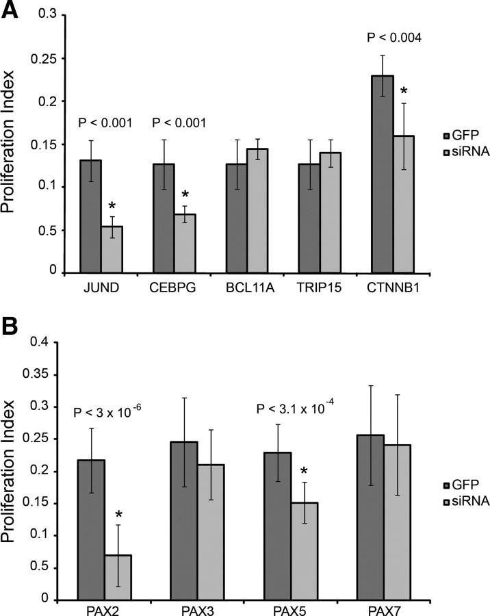
Figure 3.
Effects of siRNA treatments on SC proliferation. Proliferation phenotypes were quantified for each siRNA knockdown compared to a GFP control by calculating a proliferation index. BrdU-labeled proliferating cells were compared to the total number of DAPI-stained cells to calculate a percentage proliferation for genes differentially expressed during SE regeneration (A) and PAX genes that were upregulated during SE regeneration (B).

