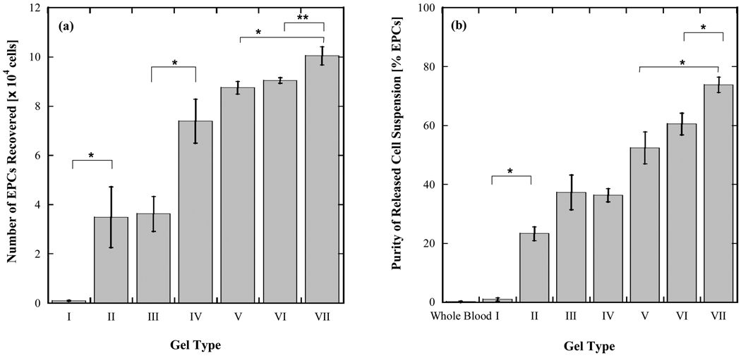
Figure 4.
Yield (a) and purity (b) of EPCs captured from whole blood within microfluidic devices coated with PEG- and antibody-functionalized hydrogels. 300 µL of whole blood collected in heparin tubes was directly injected into individual microfluidic devices and 10 devices were run in parallel. Cells released from each device were pooled into a single suspension to allow enumeration by flow cytometry. Data reported in (a) and (b) represent yield and purity for EPCs recovered from a total blood volume of 3 mL. Error bars denote standard deviations based on 3 independent measurements of EPC and total cell counts made with the same sample.

