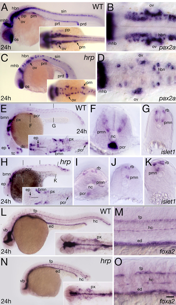
Fig. 4. Neural patterning defects in harpy mutants. Panels show wild-type reference embryos and harpy mutants.
- (A, C) Whole mount side views; insets show dorsal views of head-trunk region.
- (B, D) High resolution dorsal views of hindbrain.
- (E, H) Whole mount side views; insets show dorsal views of head region. The midline of the CNS (black line) is indicated near the epiphysis.
- (F,G, I–K) Transverse sections at levels indicated in whole mounts. Note lacking primary motor neurons, normally a bilaterally paired structure.
- (L, N) Show a whole mount side view; insets show dorsal views of head region.
- (M, O) Show high resolution side views of trunk. Note the gaps between the large cells of the mutant floor plate and hypochord.
Abbr:
bmn, branchio-motor nuclei;
dc, diencephalon;
ep, epiphysis;
ed, endoderm;
fp, floor plate;
hbn, hindbrain neurons;
hc, hypochord;
mhb, midbrain-hindbrain border;
nc, notochord;
os, optic stalk;
ov, otic vesicle;
pcr, pancreas;
pp, pharyngeal pouch;
pmn, primary motor neurons;
prn pronephric neck;
prt pronephric tubule;
prd, pronephric duct;
px, pharynx;
rb, Rohon-Beard sensory neuron;
scn, spinal cord neurons.
tc, telencephalon;
vb, ventral brain;
Scale bar = 100 µm (A,C,E,H,L,N), 40 µm (B,D,F,G,I,J,K,M,O).

