
Figure 4.
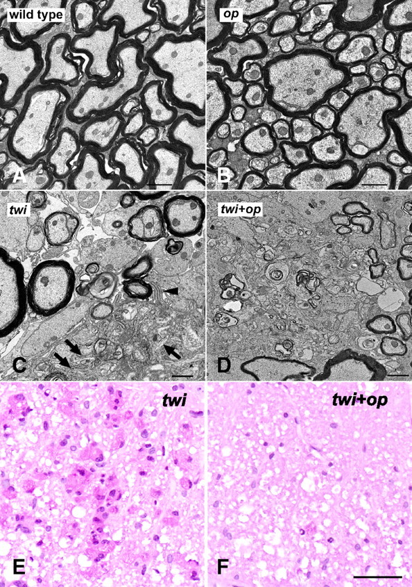
Myelin phagocytosis is compromised in the twi+op mutant. A–D, Representative electron micrographs from the ventral white matter of the cervical spinal cord demonstrate that myelination in op mice was comparable to that in wild-type mice (A, B), myelin debris was actively cleared by a macrophage (occupying lower right corner) showing pseudopodia (arrowhead) and tubular inclusion bodies (arrows) (C), and that nonphagocytosed myelin debris (arrows) was frequently observed in the white matter parenchyma of twi+op mice (D). E, F, PAS staining in the ventral white matter of the cervical spinal cord shows numerous macrophages that contain PAS-positive material in twi mice at P45 (E), whereas the numbers are few (arrowhead) in twi+op mice (F). Scale bars: A–D,1 μm; E, F, 50 μm.