
Figure 6.
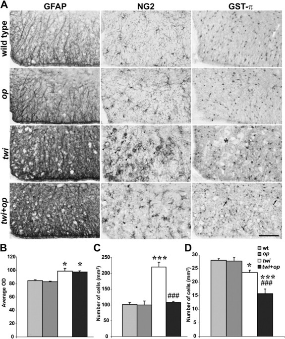
An increase in the number of OPCs in twi mice is suppressed in twi+op mice. A, Immunohistochemical demonstration of the markers for astrocytes (GFAP), OPCs (NG2), and mature OLs (GST-π) in the ventral white matter of the cervical spinal cord at 45 d old. Some macrophages were also NG2+ in twi mice (arrowheads). The asterisk shows a patchy loss of GST-π+ OLs in twi mice. Arrows indicate typical GST-π+ OLs in twi+op mice. B–D, Quantitative analyses of average ODS of GFAP immunoreactivity (B), the number of NG2+ OPCs (macrophage-like cells without processes were excluded) (C), and the number of GST-π+ OLs (D) in the ventral white matter of the spinal cord as shown in A. One-way ANOVA was followed by Scheffé's F test; *p < 0.05 and ***p < 0.001, compared with wild-type mice, and ###p < 0.001 compared with twi mice. Scale bar, 100 μm.