
Figure 9.
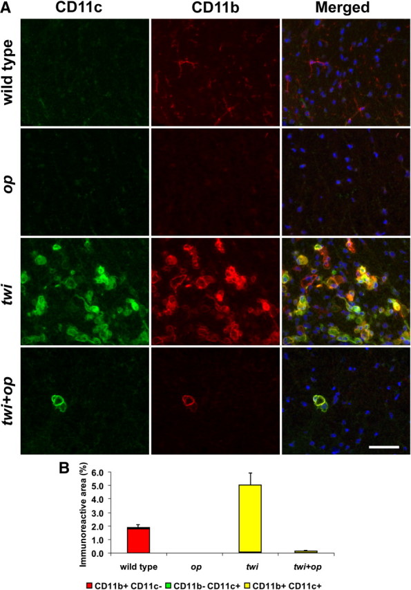
Most macrophages express CD11c antigen in twi and twi+op mice. A, Double-immunofluorescence staining for CD11c and CD11b in the ventral white matter of the spinal cord at P45. CD11b+ resting microglia in wild-type mice were mostly CD11c−, whereas the representative area with densely accumulated macrophages in twi shows that most CD11b+ cells were also CD11c+. There were few macrophages in twi+op mice, but they were mostly positive for CD11c as well. B, Morphometric analysis of the CD11c and/or CD11b-immunoreactive areas as shown in A. Error bars represent SEM for the sum of CD11b+/−CD11c+/− areas per group (n = 4). The merged images include 4′,6-diamidino-2-phenylindole (DAPI) counterstaining in blue. Scale bars, 50 μm.