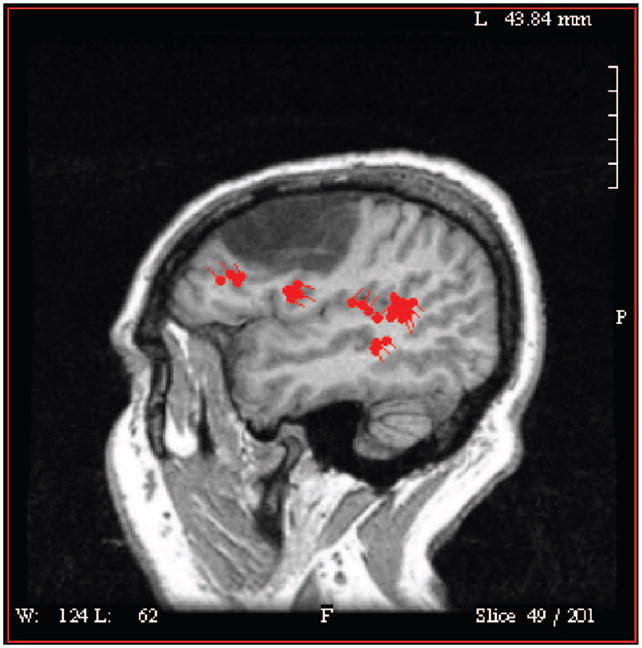
Figure.

This figure represents the regions of activation (red areas) during a word recognition test using MEG localization in a subject who suffered a left hemisphere stroke (hypodense region) and resulting aphasia. Both the primary auditory cortex and regions anterior and medial to the infarction show activation demonstrating plasticity of the brain response.
