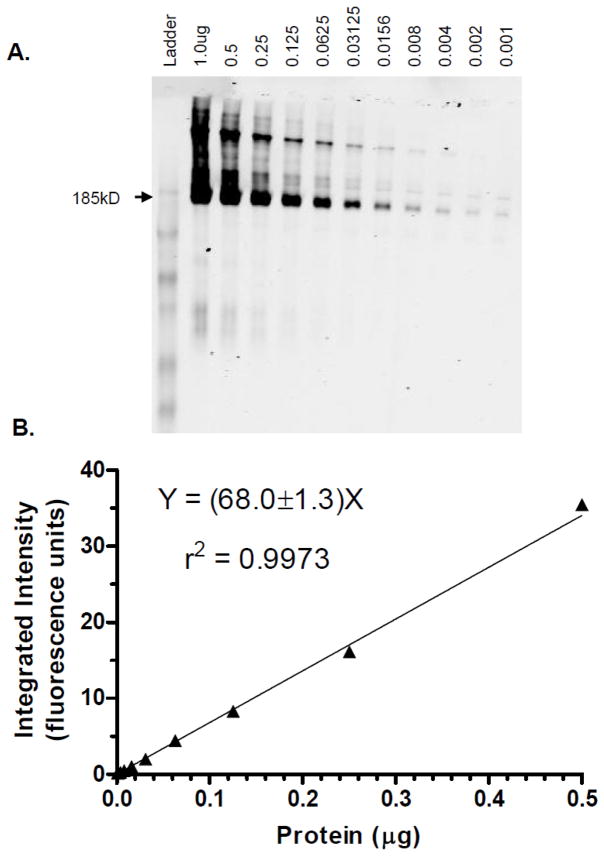
Figure 1.
Representative immunofluorescent blotting results for membrane vesicles isolated from MRP2-overexpressing Sf9 insect cells. A: Scanned image of blot converted to black and white; Sf9 cells lacking MRP2 are non-immunoreactive (data not shown); protein amounts (μg) as shown. B: Corresponding calibration curve weighted 1/Y, with linear regression results as shown.

