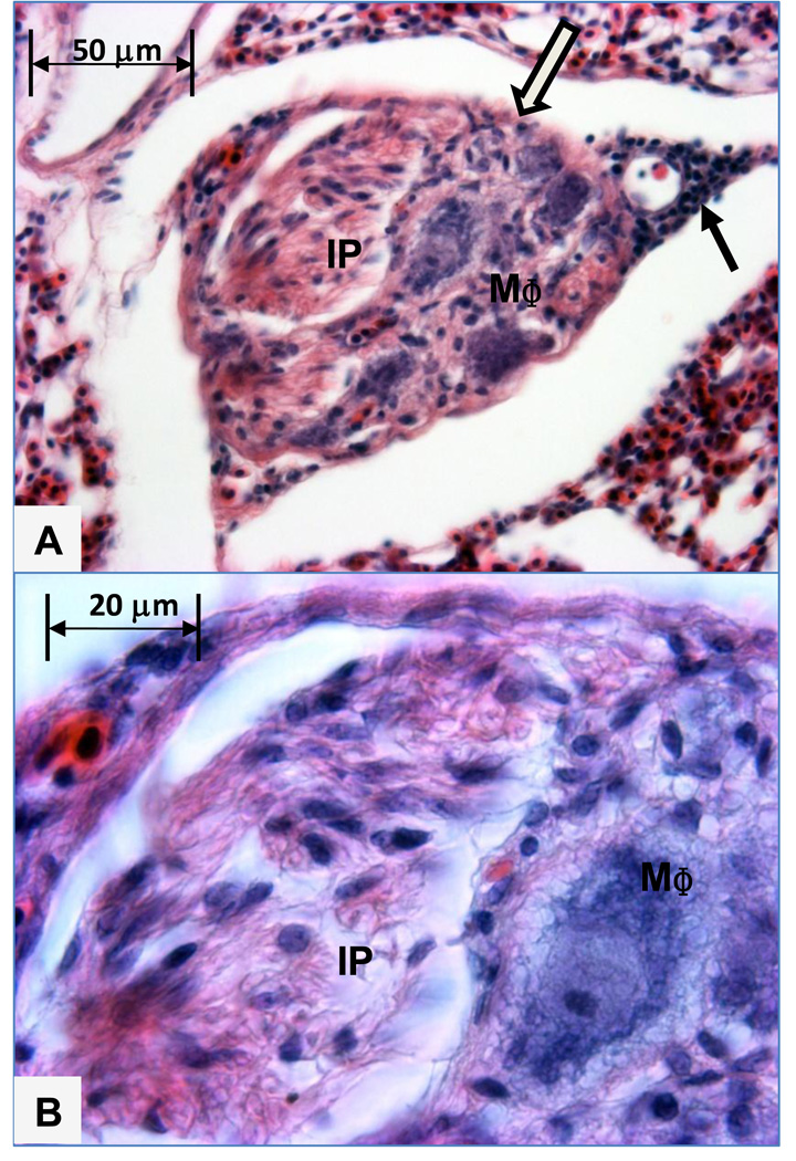
Figure 4.
A. This intermediate sized plexiform lesion developed in an interparabronchial arteriole branch coursing within the septum between adjacent parabronchi. The lesion contains foam-type macrophages (MΦ) adjacent to the remnant of the arteriole wall on the bottom right and a channel filled intimal proliferating cells (IP) adjacent to the arteriole smooth muscle (open arrow) on the upper left. Filled arrow indicates perivascular mononuclear cells outside the wall of the arteriole. Figure 4B. Higher magnification shows the intimal proliferating cells (IP) and a foam-type macrophage (MΦ).

