
Fig. 4.
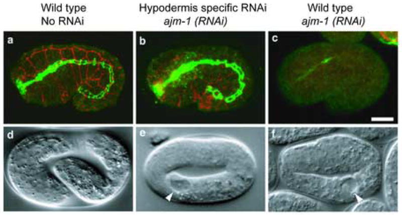
Tissue-specific depletion of AJM-1 via ajm-1(RNAi) in wild type and hypodermis-rescued rde-1 embryos.
a–c, MH27 (a monoclonal antibody specific to AJM-1) staining of wild type, ajm-1(RNAi); rde-1 [plin-26::rde-1] and ajm-1(RNAi) embryos. Hypodermal MH27 signal is shown in red (a projection of three confocal sections of the surface of the embryo), and pharyngeal and intestinal signal is shown in green (a projection of two central confocal sections). In the wild type, MH27 staining is bright and even in all epithelia (a). ajm-1(RNAi) in the strain rescued for rde-1 in the hypodermis (b) results in marked reduction of MH27 staining in the hypodermis, but no effect in the pharynx and intestine. ajm-1(RNAi) in a wild-type embryo causes almost complete loss of MH27 signal from all embryonic epithelia (c). d–f, Nomarski phenotypes of wild type, ajm-1(RNAi); rde-1 [plin-26::rde-1] and ajm-1(RNAi) embryos. A wild type embryo is shown at the 3-fold stage (d). ajm-1(RNAi) causes a nearly identical 2-fold arrest phenotype in both the strain rescued for rde-1 in hypodermis (e) and in the wild type (f). However, in the case of the strain rescued for rde-1 in hypodermis, embryonic lethality is only observed in 50% of the embryos, while 100% lethality is observed in a wild-type background. Scale bar represents 10 μm. Arrowheads indicate vacuoles characteristic of the ajm-1 loss of function phenotype.