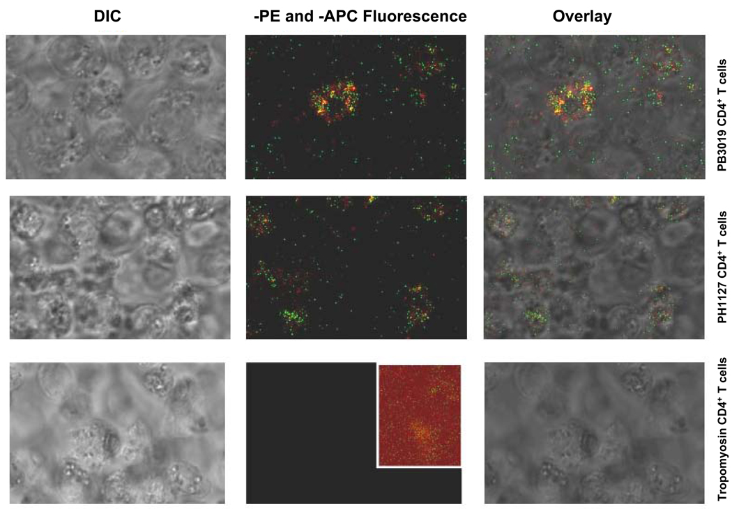
Figure 5. NS3358–375 and S370P MHC class II tetramers are able to bind to CD4+ NS3358–375 T cells.
The fluorescent images were all taken using identical confocal settings including laser power, emission filters, pinhole, and scan speed. NS3358–375 specific CD4+ T cells (Materials and Methods) were stained with both NS3358–375-PE (green) and S370PAPC (red) MHC class II tetramers at 10µg/ml. NS3358–375 specific CD4+ T cells from PB3019 and PH1127 are double positive for NS3358–375 and S370P tetramers (rows one and two). CD4+ Tropomyosin-specific T cells were used as a control (third row). The inset (row 3, panel 2) has been multiplied by three to reveal the level of background for comparison to the experimental panels shown in the same column.

