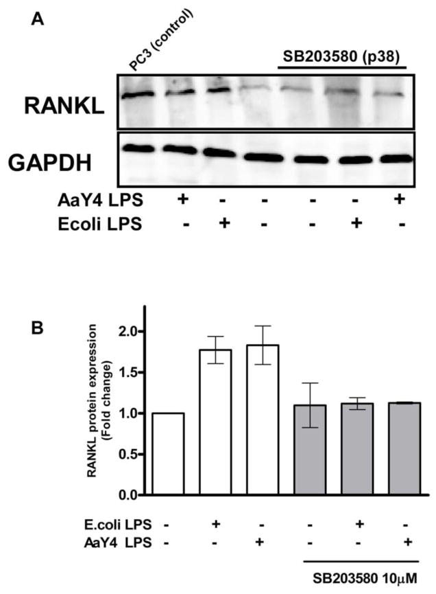
Figure 3. RANKL protein expression by LPS-stimulated PDL cells is dependent on p38 MAPK activity.
PDL cells grown on 6-well plates were de-induced for 12 hours in culture medium containing 0.3% FBS and then stimulated with LPS from either E.coli or A. actinomycetemcomitans for 72 hours. The specific inhibitor for p38 MAPK (SB203580; 10 μM) was added to the culture medium 30 minutes before stimulation with LPS (1 μg/mL). (A) Western blot analysis of RANKL expression from PDL whole cell lysates. Positive control for RANKL expression is cell lysates from the human prostate cancer cell line (PC-3). (B) Density analysis of data from three independent experiments shows significant inhibition of LPS-induced RANKL protein by SB203580 (* p<0.05).

