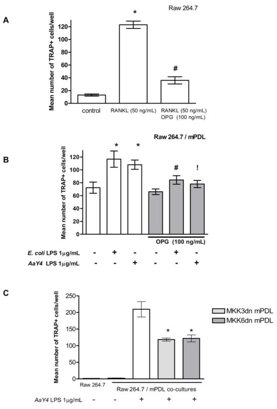
Figure 4. LPS-stimulated PDL cells stimulate osteoclastogenesis, which is regulated by p38 MAPK pathway.
Stimulation with RANKL induces RAW 264.7 cells to differentiate into multinucleated TRAP+ cells, whereas pre-treatment with OPG inhibits this effect *Indicates significant (p<0.05) difference from unstimulated cells and #indicates a significant decrease on the number of osteoclasts with OPG treatment (A). mPDL cells were stimulated with LPS from E. coli and A. actinomycetemcomitans Y4 and cultured 3 days. These cells were then co-cultured with RAW 264.7 cells for an additional 6 days and the number of multinucleated, TRAP+ cells was counted. Stimulation with LPS increased significantly the number of TRAP+ cells and this effect was inhibited by OPG *Indicates significant (p<0.05) difference from unstimulated cells and #indicates a significant decrease on the number of osteoclasts with OPG treatment. !Indicates p=0.056 for the significance of the decrease on the number of osteoclasts with OPG treatment in A. actinomycetemcomitans Y4 LPS-stimulated cells. (B). Co-culture of RAW cells with stable PDL cell lines over-expressing dominant negative mutants of MKK3 (MKK3dn-PDL) and MKK6 (MKK6dn-PDL), upstream activators of p38 MAPK, significantly decreased the number of TRAP+ cells *Indicates significant (p<0.05) difference from non-transfected PDL cells (C). Bar graphs indicate mean ± standard deviation of number of TRAP+, multinucleated cells counted in each well.

