
Fig. 4.
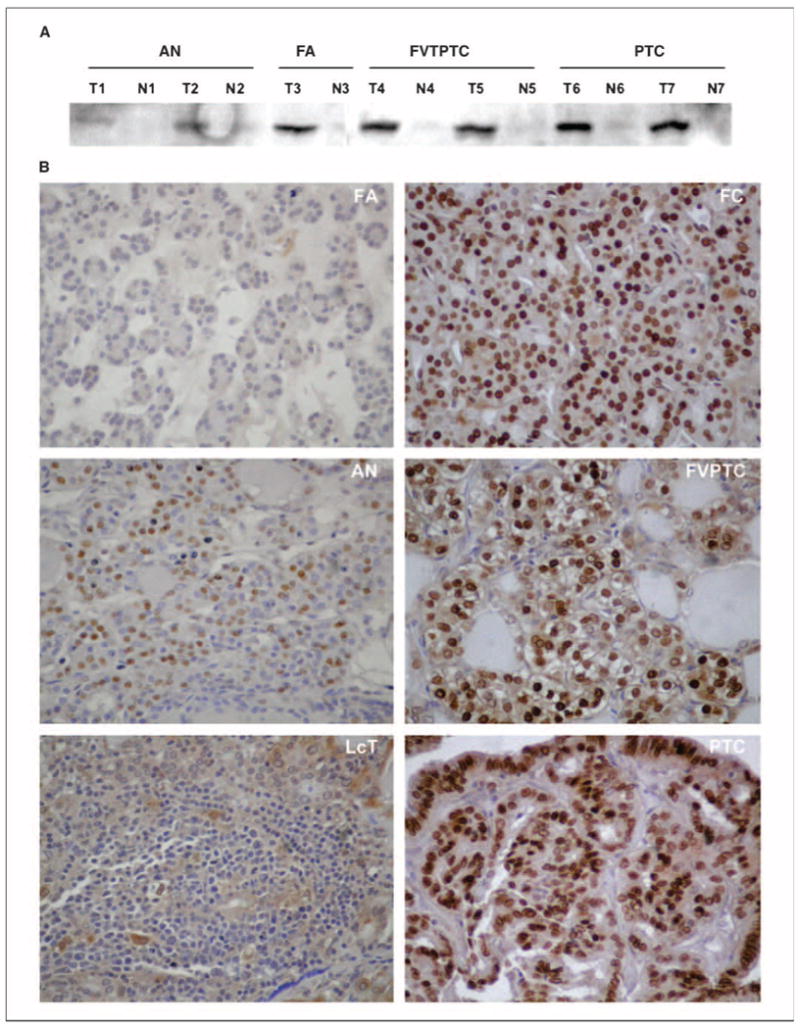
HMGA2 expression in thyroid. A, Western blot analysis of HMGA2 protein expression in thyroid tumors (T1-T7) and in adjacent normal thyroid tissues (N1-N7).The anti-HMGA2 goat polyclonal antibody recognized HMGA2 expression specifically in thyroid tumors but not in adjacent normal thyroid tissue. High protein expression of HMGA2 is detected in malignant tumors (T4,T5,T6,T7) compared with benign tumors (T1,T2,T3). B, immunohistochemistry of HMGA2 in thyroid tumors. Note the intense nuclear staining of most nuclei within the malignant tumors compared with low or negative expression in the benign tumors. No detectable expression was seen in the adjacent normal thyroid tissue. Magnification, 400×.