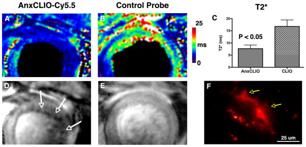
FIGURE 4.
Sensitivity of AnxCLIO-Cy5.5 for cardiomyocyte (CM) apoptosis.16 Postpartum Gaq-overexpressing mice with heart failure have been used. These mice develop a postpartum cardiomyopathy with very low levels of apoptosis (1–2%), minimal inflammation and necrosis, and normal capillary membrane permeability. AnxCLIO-Cy5.5, however, is able to penetrate the interstitial space and detect the very sparsely expressed apoptotic CMs. Mice injected with the active probe are shown in (a, d) and those injected with the control probe in (b, e). T2* maps are shown in (a, b) and T2*-weighted images in (d, e). T2* is significantly reduced in the animals injected with AnxCLIO-Cy5.5 and numerous discrete foci of probe uptake (signal hypointensity, white arrows) are seen. (f) Fluorescence microscopy of a mouse injected with AnxCLIO-Cy5.5 shows the uptake of the agent by an apoptotic CM with characteristic membrane blebbing (yellow arrows) (Reprinted with permission from Ref 16. Copyright 2009 the American Heart Association).

