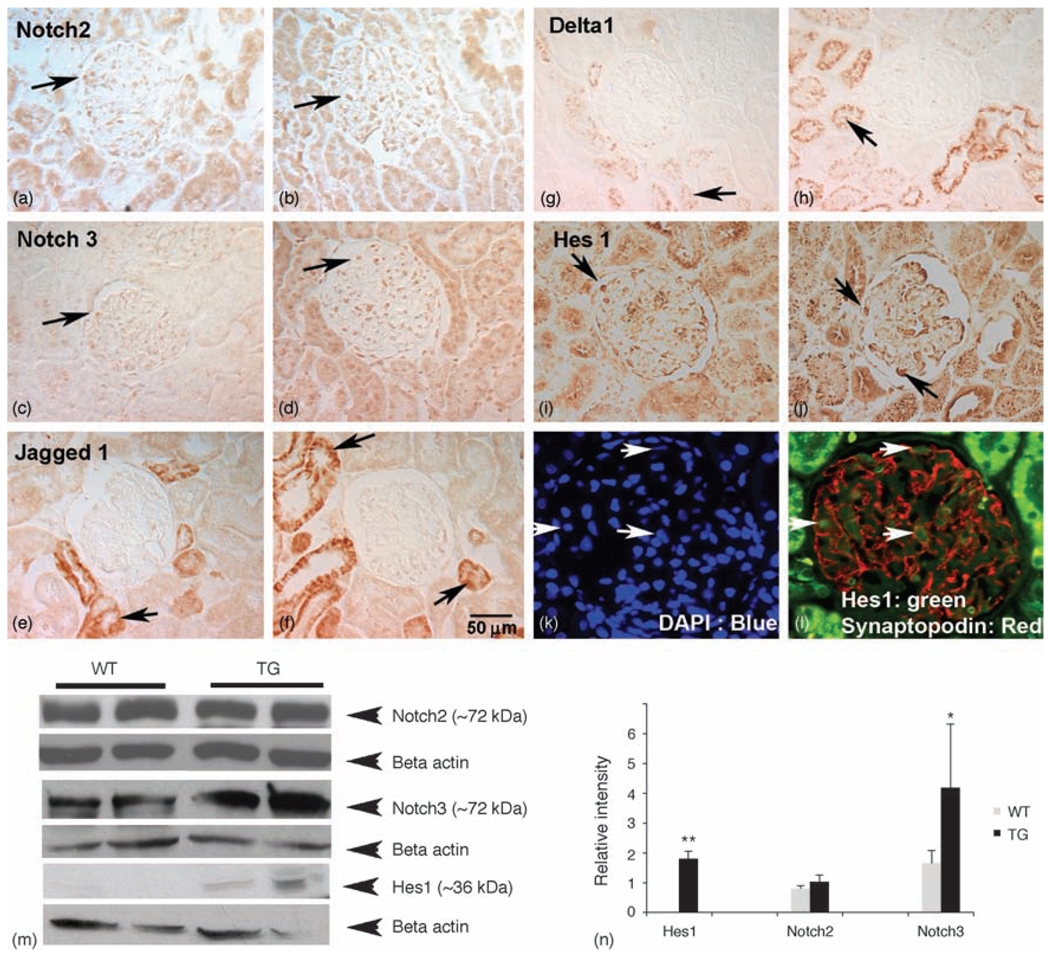
Fig. 2. Upregulation of Notch pathway members in HIV-transgenic rat kidneys.
Kidney sections were labeled for Notch2 IC, Notch3 IC, Jagged1, delta1 and Hes1 expression. (a and b) Notch2 intracellular was expressed in both glomeruli and tubules from WT (a) and HIV-Tg rat sections (b). (c and d) Notch3 IC was upregulated in the HIV-Tg kidneys in both tubular and glomerular cells (d) compared with WT kidney sections (c). (e and f) Jagged1 was not expressed in the glomeruli; however, more tubules from HIV-Tg rats (f) expressed Jagged1 compared with WT controls (e). (g and h) Similar to Jagged1, delta1 was not expressed in glomeruli; however, expression was increased in tubules (arrow in h) in HIV-Tg sections compared with the WT controls (g). (i and j) Hes1 was minimally expressed in the WT kidneys (i), but was upregulated in the glomeruli from HIV-Tg kidneys (arrows in j). (k and l) To identify whether Hes1-positive cells are podocytes, double immunoflourescence was performed using antibodies specific for synaptopodin (red) and Hes1 (green), and counterstained with DAPI (k). Arrows indicate cells double positive for synaptopodin and Hes1 (l). All figures except panels (k) and (l) are original magnification 40×. Panels (k) and (l) are magnified from 40×. (m and n) Protein lysates prepared from WT and TG kidneys were subjected to western blot analysis for quantitation of Notch2 IC, Notch3 IC and Hes1. Notch2 IC did not vary between WT and TG samples, whereas Notch3 IC and Hes1 were increased in TG kidney lysates compared with WT lysates. Densitometric analysis of western blots from at least three experiments shows an increase in Hes1 and Notch3 expression in HIV-Tg kidneys. DAPI, 4′-6-Diamidino-2-phenylindole; Hes1, hairy enhancer of split homolog-1; HIV-Tg, HIV-transgenic; IC, intracellular; TG, HIV-Tg; WT, wild-type.

