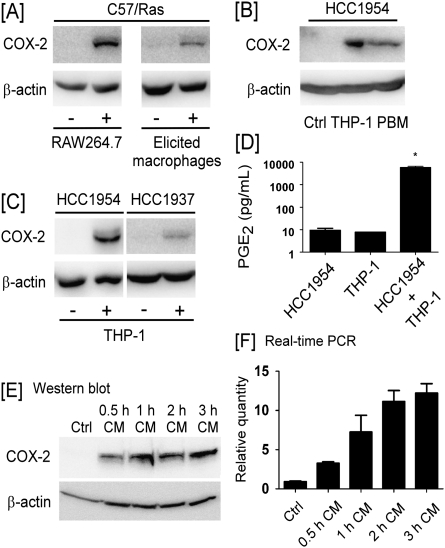
Fig. 1.
Macrophages induce COX-2 expression in breast cancer cells. (A) C57/Ras cells were cocultured (24 h) with RAW264.7 macrophages or elicited peritoneal macrophages. (B) HCC1954 cells were cocultured (24 h) with PMA-treated THP-1 monocytes or IFNγ/lipopolysaccharide-activated PBMs. (C) HCC1954 or HCC1937 cells were cocultured (24 h) with THP-1 cells. Western blotting was utilized to determine levels of COX-2 and β-actin in cell lysates. (D) PGE2 levels in CM recovered from HCC1954 cells, THP-1 cells and cocultured cells were measured utilizing enzyme-linked immunosorbent assay. Data are mean ± standard deviation (n = 3; *P < 0.05). (E and F), CM from HCC1954 and THP-1 cells cocultured for 0.5–3 h were added to naive HCC1954 cells and incubated for 6 h. COX-2 expression was monitored utilizing (E) western blot analysis and (F) quantitative real-time–PCR. The real-time PCR values of COX-2 are normalized to β-actin, and the relative values are shown as mean ± standard deviation (n = 3).

