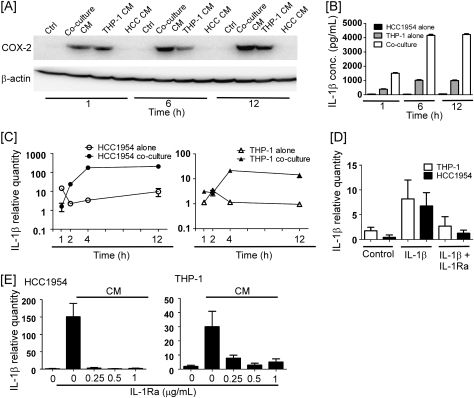
Fig. 6.
Autoamplification led to increased production of IL-1β in macrophages cocultured with breast cancer cells. (A) HCC1954 and THP-1 cells were cultured alone or cocultured for the indicated time periods. CM collected at each time point were then used to treat naïve HCC1954 cells for 6 h. COX-2 and β-actin levels in HCC1954 lysates were determined by western blot. (B) HCC1954 and THP-1 cells were cultured alone or cocultured for the indicated time periods. IL-1β concentrations in CM were determined utilizing enzyme-linked immunosorbent assay. Data are mean ± standard deviation (n = 3). (C) HCC1954 and THP-1 cells were cultured alone or cocultured for the indicated time periods. (D) HCC1954 and THP-1 cells were treated with vehicle, IL-1β (10 ng/ml) or IL-1β and IL-1Ra (0.5 μg/ml) for 3 h. (E) Naive HCC1954 and THP-1 cells were treated with 1 h coculture CM in the absence or presence of the indicated concentrations of IL-1Ra for 3 h. In panels C–E, total RNA was isolated, and quantitative real-time PCR was performed. Levels of IL-1β mRNA were normalized to levels of β-actin mRNA. Data are mean ± standard deviation (n = 3).

