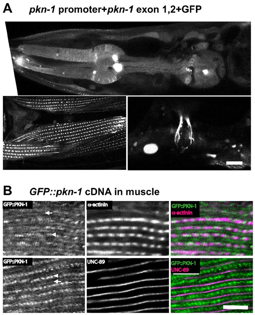
Figure 2. The expression and localization of PKN-1 in muscle.
A: GFP expressed by the pkn-1 promoter was observed in many types of muscle. We injected a mixture of pKK44 plasmid (5 Kb of upstream putative promoter, the first and second exons, and the first intron of pkn-1 was cloned into pPD95.75, a gift from Dr. Fire) and a lin-15 rescuing plasmid into lin-15 (n765) (temperature sensitive multinulva (Muv)) worms. To obtain transgenic animals, we picked non-Muv worms at 25°C. In transgenic animals, GFP driven by pkn-1 promoter was observed in pharynx including pharyngeal muscle (upper panel), body wall muscle (lower left panel), and vulva muscle (lower right panel). Scale bar, 10 µm.
B: GFP tagged PKN-1 is localized at dense bodies (arrowheads) and M-lines (arrows). We prepared transgenic worms harboring an extrachromosomal array containing both a plasmid expressing GFP tagged full-length pkn-1 cDNA under the control of heat shock promoter, and a plasmid for the rol-6 (roller) dominant marker. Worms were fixed by the Nonet method29. Antibody staining with anti-GFP (Invitrogen A11122, 1/200 dilution), and MH35 (1/200 dilution; α-actinin) or MH42 (1/200 dilution; UNC-89) was performed as described previously28. All images were captured at room temperature with a Zeiss confocal system (LSM510) equipped with an Axiovert 100M microscope using an Apochromat 63x/1.4 oil objective, in 2.5x zoom mode. The color balances of the images were adjusted by using Adobe Photoshop. Scale bar, 10 µm.

