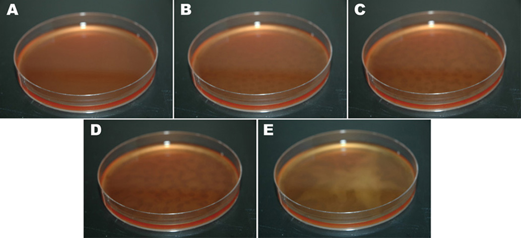
Fig. 7.
Observation of rosette swarming in Leishmania. A, 0 h; B, 2 h; C, 4 h; D, 8 h; E, 30 h. At 0 h, a confluent cell suspension is observable. At 2 h, a spotty appearance is detectable corresponding to regions of higher and lower cell density. These elongate at 4 h and begin to converge at 8 h. Cells eventually congregate into a star-like appearance in the center of the Petri dish.

