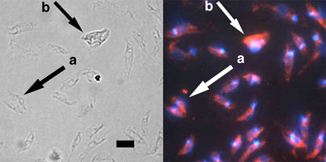
Fig. 9.
Labeling of fusion bodies by Leishmania specific anti–gp63 monoclonal antibody (mAb). Left panel, light microscopy; right panel, fluorescence microscopy. Numerous individual promastigotes are visible and contain a single nucleus and single kinetoplast stained with DAPI. A dividing cell (arrow a) and fusion body (arrow b) are shown along with individual promatigotes. All cells including the fusion body were detected by the Leishmania specific anti–gp63 mAb, demonstrating that fusion bodies are not artifacts or contaminants. Fusion bodies contain multiple DAPI staining foci as expected by the fusion of two or more leishmanial promastigotes. The ratio of the integrated pixel density of DAPI DNA staining in the dividing cell and fusion body compared to five individual promastigotes in the same micrograph were 1.88 and 2.70, respectively. Scale bar, 10 µm.

