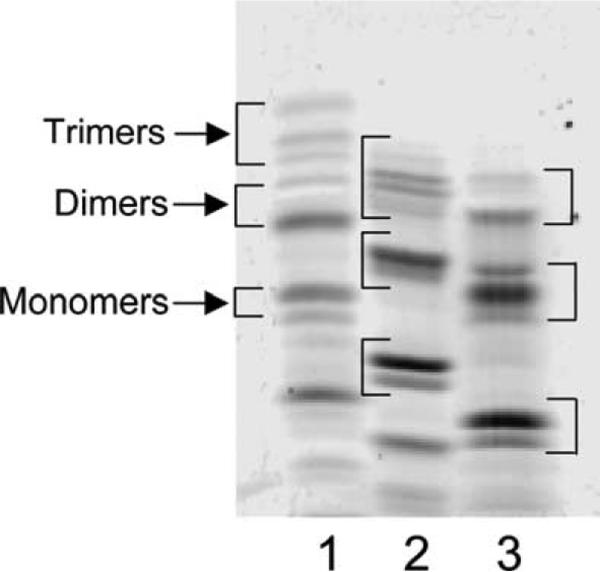
Fig. 2.
Muropeptides labeled with different fluorophores. E. coli CS109 peptidoglycan was digested with N-acetylmuramidase, and the resulting muropeptides were labeled with the fluorophores, ANSA (lane 1), ANDA (lane 2), and ANTS (lane 3). The samples were separated by using the optimal conditions established for FACE gel electrophoresis as described under Materials and methods. The brackets in each lane denote the approximate positions of trimers, dimers, and monomers, respectively. Monomer muropeptides are composed of a single N-acetylglucosamine-N-acetylmuramic acid (NAG–NAM) glycan subunit plus an amino acid side chain of two to five residues, dimer muropeptides are composed of two NAG–NAM glycan subunits cross-linked to one another via their peptide side chains, and trimer muropeptides are composed of three NAG–NAM glycan subunits cross-linked to one another via their peptide side chains. Bands migrating faster than monomers are low-molecular-mass constituents of the fluorescent dye solutions.

