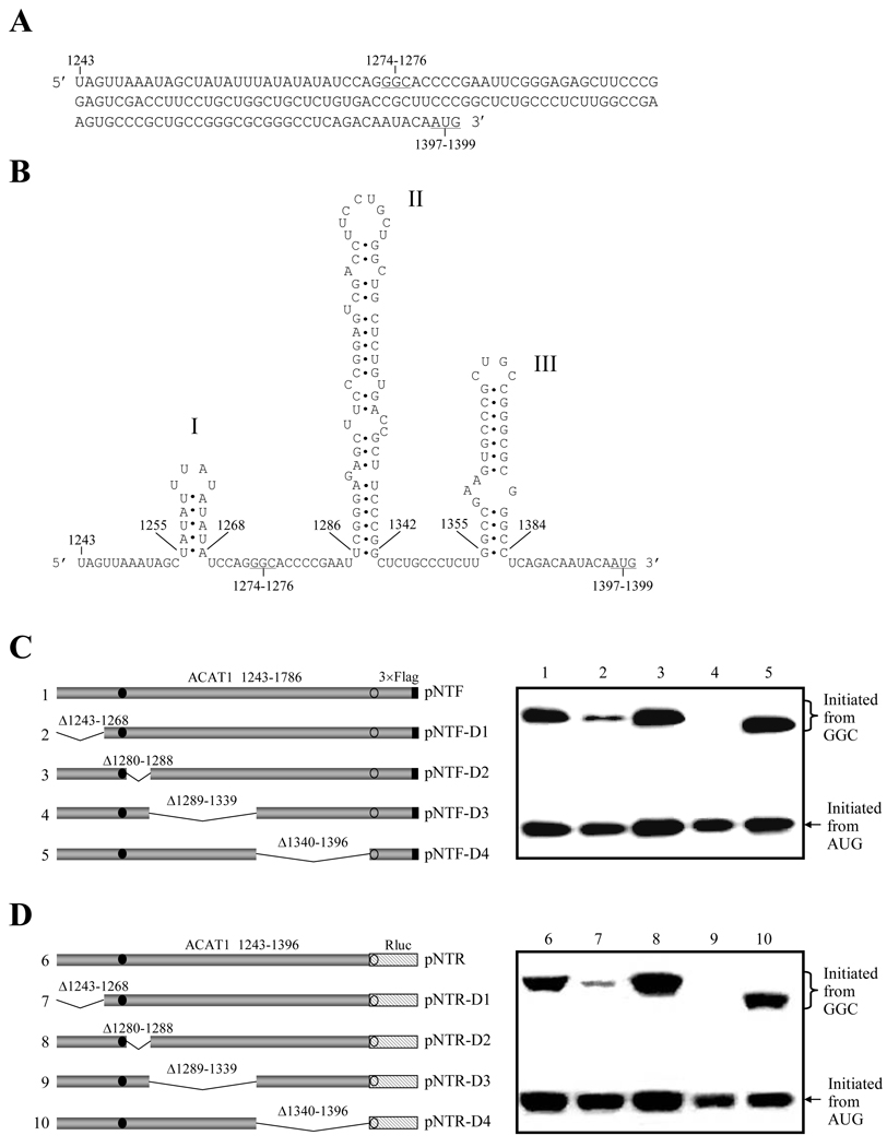
Figure 1. Predicted stem-loops in the vicinity of GGC1274–1276 codon are required for the production of ACAT1 isoforms.
(A) Vicinal sequence of the GGC1274–1276 codon (nt 1243–1396). The initiation codons GGC1274–1276 for the 56-kD human ACAT1 isoform and AUG1397–1399 for the 50-kD one are underlined.
(B) Predicted RNA secondary structures in the vicinity of GGC1274–1276 codon (nt 1243–1396). The three successive stem-loops are respectively labeled with I, II and III.
(C) Schematic representation of the partial ACAT1 mRNA sequence (nt 1243–1786) and its truncated forms on the left panel. The deleted regions (Δ1243–1268, Δ1280–1288, Δ1289–1339 and Δ1340–1369) are marked on the top of each bar. Gray bar, ACAT1 mRNA sequence (ACAT1 1243–1786); black bar, 3×Flag coding sequence (3×Flag); filled circle, GGC1274–1276 initiation codon; hollow circle, AUG1397–1399 initiation codon. The expression plasmids depicted on left are transiently transfected into AC29, the lysates are prepared and immunoblotting is carried out with anti-ACAT1 antibodies (DM10). The curly bracket and the arrow indicate the positions of ACAT1-NT-Flag proteins respectively initiated from GGC1274–1276 and AUG1397–1399. The experiments are repeated three times with similar results.
(D) Schematic representation of the replacement of partial ACAT1 AUG-ORF with the whole Renilla luciferase AUG-ORF on the left panel. Gray bar, vicinity of GGC1274–1276 codon (ACAT1 1243–1396) (ACAT1 1243–1396); hatched bar, the whole AUG-ORF of Renilla luciferase (Rluc); others representing the same in (C). The expression plasmids depicted on left are transiently transfected into AC29, the lysates are prepared and immunoblotting is carried out with anti-Rluc antibody. The curly bracket and the arrow indicate the positions of the fused ACAT1-Rluc protein initiated from the GGC1274–1276 and Rluc protein initiated from AUG1397–1399. The experiments are repeated three times with similar results.

