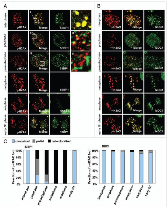
Figure 1.
The components of IRIF are reorganized during the cell cycle. HeLa cells were synchronized by colcemid, exposed to 1 Gy IR and fixed after 30 min incubation. Mitotic stages were classified by DAPI staining. (A) Immunostaining for γH2AX and 53BP1. I-III refer to enlargements of the noted areas. (B) Immunostaining for γH2AX and MDC1. (C) Fraction of γH2AX foci colocalized with 53BP1 (left) or MDC1 (right).

