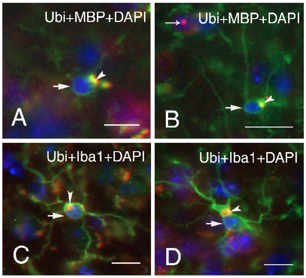
Figure 3.
Representative immunofluorescent staining (arrows) of oligodendrocytes of wildtype (A) and CGG KI (B) mice, and microglia of wildtype (C) and CGG KI mice (D). Large, irregularly shaped cytoplasmic inclusion bodies were found in oligodendrocytes and microglia of both wildtype and CGG KI mice (arrow heads). Oligodendrocytes were labeled for myelin basic protein (MBP, green) and microglia for Iba1 (green), so that co-colocalization of ubiquitin in the inclusions with either MBP or Iba1 appears yellow. Nuclei were stained with DAPI (blue). Both wildtype mice were 60 weeks of age (A, C), and the two CGG KI mice were 70 (B) and 59 (D) weeks of age. Scale bars are 10 µm.

