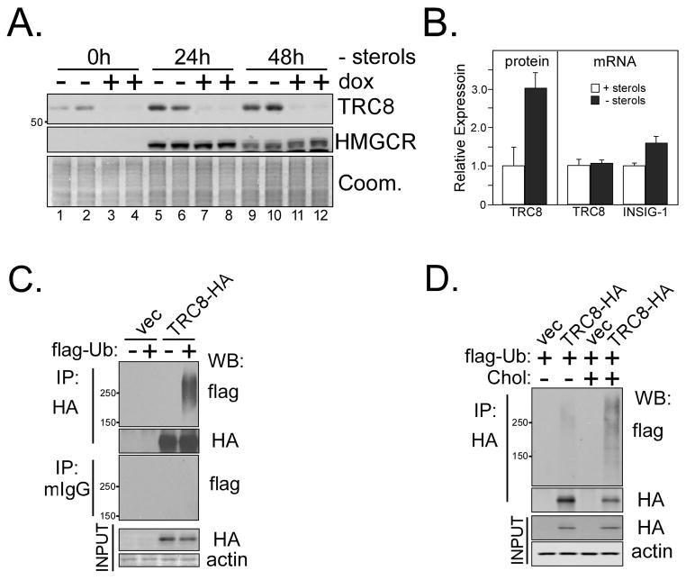
Figure 2. Sterols regulate endogenous TRC8 and induce ubiquitylation.
(A) Membranes purified from HEK293 cells stably transfected with a tet-inducible shRNA targeting TRC8 permitted detection of endogenous TRC8 by Western blot (lanes 1, 2, −dox). Identity of the band was confirmed by 4 days of dox treatment (+dox) to induce knockdown. Parallel cultures were sterol-starved for 24 (lanes 5–8) or 48h (lanes 9–12) and membrane preparations analyzed for endogenous TRC8 and HMGCR (postive control). Coomassie staining verified equal loading. (B) Data for 24 h sterol starvation were densitized and quantified. Each bar represents the mean of TRC8 signal in triplicate samples, +/− s.d. RNA isolated from the same cultures was analyzed by realtime RT-PCR for endogenous TRC8 and INSIG-1 mRNAs. Delta Ct values were converted to fold expression and normalized to control samples (+sterols). (C) TRC8-HA or control (vec) HEK293 cells, grown in sterol-replete medium, were transfected with flag-tagged ubiquitin (1 μg), dox-induced and harvested 24 h later following MG132 addition (10 μM) during the final 2h. Lysates (200 μg aliquots) were immunoprecipitated with anti-HA or murine IgG (controls) and IP pellets analyzed on Western blots for flag-ubiquitin. HA immunoblots showed equal recovery of TRC8-HA; this and other HA blots were cropped to remove strong IgG background. Actin and HA blots verified equal input. (D) TRC8-HA and control cells were transfected with flag-ubiquitin and induced with dox as in (C). Cells were then sterol-starved for 15h followed by addition of cholesterol (12.5 μM) and 25-HC (10 μg/mL) for 3h to the indicated plates. MG132 was added 2h prior to harvest and lysates (200 μg aliquots) were immunoprecipated with anti- HA beads. Western blot analysis utilized the indicated antibodies.

