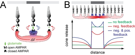
Figure 8. A conceptual model of positive feedback in the outer retina.
(A) Diagram depicting the differential spread of positive and negative feedback within an HC. The top bar denotes the illumination pattern. A cone depolarized in darkness will release glutamate, activating AMPA receptors (APMARs), causing depolarization and Ca2+ influx. The rise in Ca2+ is restricted to the specific dendrite that contacts the cone, and the resulting positive feedback is localized to that cone. The depolarization spreads electrotonically through the HC, resulting in negative feedback from all of the dendrites. (B) Model simulations of the effect of feedback on synaptic release from a linear array of cones exposed to a dark spot on a non-saturating light background (see Methods). The positive feedback signal (blue) is localized to HC dendrites in contact with dark cones while the negative feedback signal (red) electrotonically spreads through the HCs. Traces show simulated cone release with no feedback (green), with negative feedback, (red), and with equally weighted negative and positive feedback (blue).

