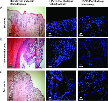
Figure 1.
Histological depiction of the rhesus macaque cervix and its infection by human papillomavirus (HPV) pseudovirus. Representative images from the three regions of the cervix are shown: A) ectocervix, B) transformation zone, and C) endocervix. Hematoxylin- and eosin-stained tissues are depicted in micrographs in the first column (scale bar = 40 μm). The images in the second and third columns are representative images of cervical specimens from monkeys challenged with HPV16-pseudovirus (PsV) transducing the red fluorescent protein without and with cytology sample collection, respectively (scale bar = 10 μm). Red stained cells indicate PsV-infected cells.

