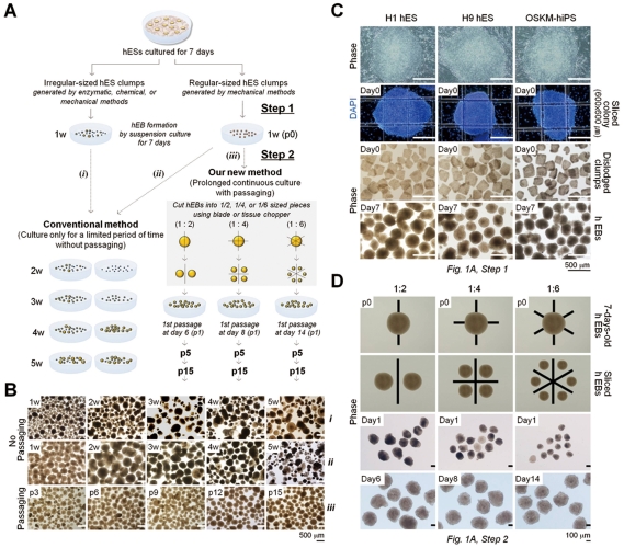
Figure 1. Long-term-cultured hEBs derived from hESCs and hiPSCs.
(A) Schematic representations of the hEB culture conditions with and without passaging. w: week, p: passage. Step 1: Period of hEB formation, Step 2: Period of hEB maintenance and expansion. (B) Morphologies of the cultured hEBs with or without passaging for various times as indicated. (i and ii) H9 hESC-derived hEBs cultured without passaging (conventional method). (iii) H9 hESC-derived hEBs cultured with passaging. For subculturing, hEBs were sliced at a ratio of 1∶4 every 7 days. The medium was changed every 3 days (i–iii). (C) Representative images of regular-sized hEB formation. (Top) Seven-day-old (7 d) hESCs (H1 and H9 hES) and OSKM-induced hiPSCs (OSKM-hiPS). (Middle top) hESC and iPSC colonies sliced into a grid pattern (600×600 µm) using a McIlwain tissue chopper prior to dislodgement. Cell nuclei were visualized with DAPI. (Middle bottom) Mechanically dislodged colonies in suspension culture. (Bottom) The 7 d-hEB population in suspension culture. (D) Representative images of the hEB passaging process. The 7 d-hEBs derived from H9 hESCs (Top) were mechanically dissociated into smaller aggregates at ratios of 1∶2, 1∶4, and 1∶6 (Middle top). (Middle bottom) Phase contrast images of passaged hEBs cultured for 1 day. (Bottom) Representative images of passaged hEBs exhibiting complete re-growth to normal size.

