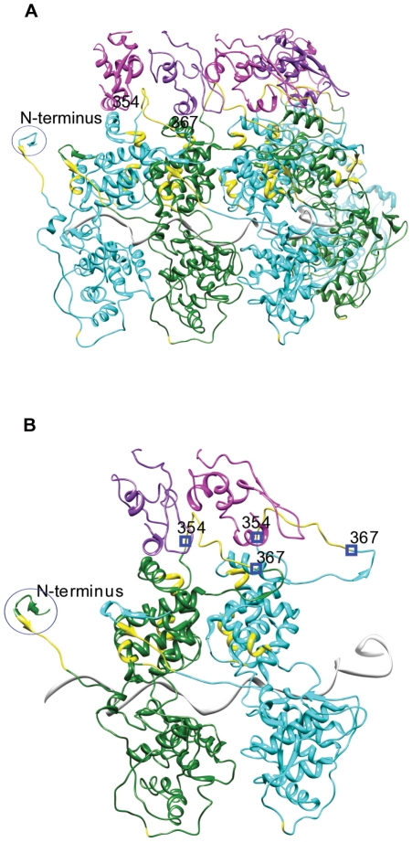
Figure 7. Crystal structure of Vesicular Stomatitis Indiana Virus nucleocapsid complexed with the phosphoprotein's nucleocapsid-binding domain(3HHW).
A.) 5 nucleoproteins colored green and cyan alternating to make them easily distinguishable and 5 nucleoprotein-binding domains of the phosphoprotein colored in magenta and purple. The predicted disordered residues are highlighted in yellow. The predicted disordered nucleoprotein residues 354–367 are shown in contact with the binding domain of the phosphoprotein. B.) Two nucleoproteins and two phosphoproteins. Chain K and L are nucleoproteins colored green and cyan. Chains A and B are phosphoproteins colored magenta and purple. The blue circle is highlighting the N-terminus of the nucleoprotein and the blue squares indicate residues 354 and 367 on each N chain. Predicted disordered residues are highlighted in yellow.

