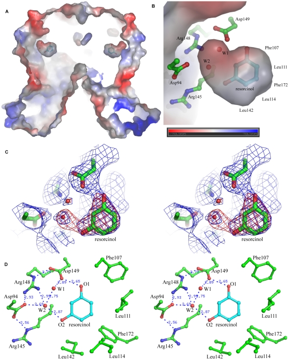
Figure 3. Recognition mode between RolR and resorcinol.
(a)The electrostatic potential map of a longitudinal section drawing of res-RolR calculated with deletion of the resorcinol molecule showing the internal binding pocket between RolR and resorcinol. (b)The section around the resorcinol of the binding pocket fitting to a resorcinol molecule and two water molecules highlighted the water-mediated interactions between resorcinol and RolR. (c) The 2Fo-Fc (blue) and Fo-Fc (red) omit map around the bound resorcinol molecule, contoured at 1σ and 3σ respectively. (d) The recognition mode between RolR and resorcinol unique in a water molecules-mediated hydrogen bond network. The sidechains of residues involved in ligand recognition and water molecules are shown in ball-and-stick, with atoms O, N and C in red, blue and green, respectively.

