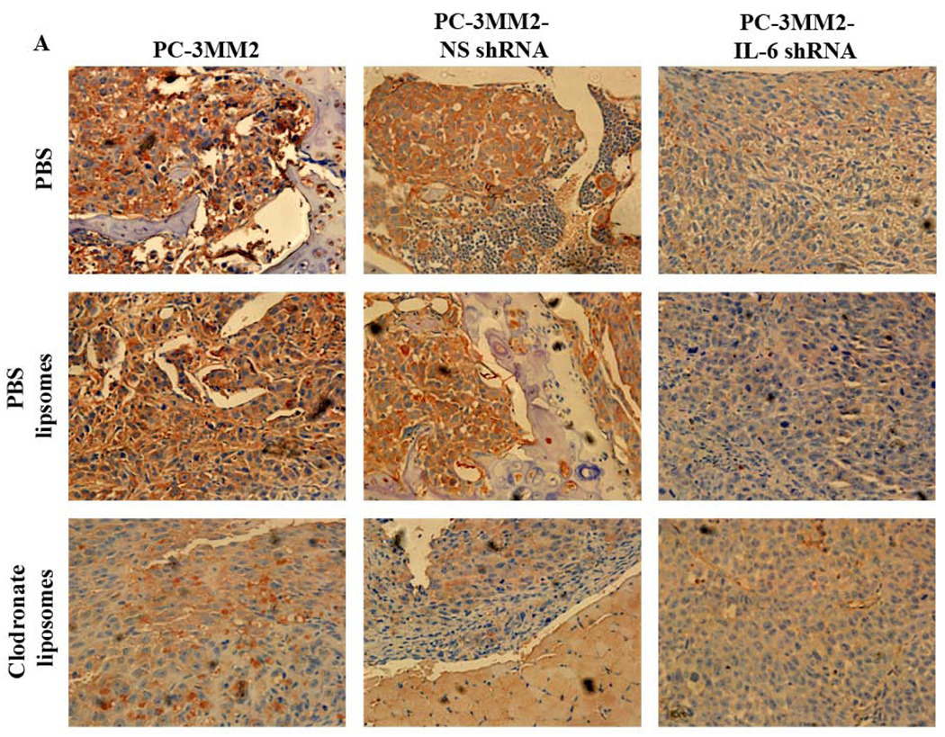
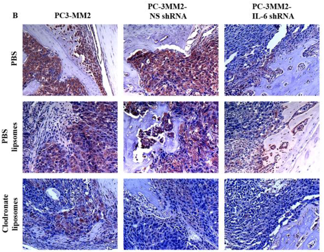
Fig. 4.
Immunohistochemical analyses. Tissues were harvested and processed for immunohistochemical analysis of the expression of IL-6 (using 3-amino-9-ethylcarbazole chromogen B) and TNF-α (using 3,3’-diaminobenzidine). A: Expression of IL-6 (red) was significantly decreased by transfection of PC-3MM2 with IL-6 shRNA. Treatment with clodronate liposomes also decreased the expression of IL-6 in bone tumors. B: Expression of TNF-α (brown) was significantly decreased by knock down of IL-6 by IL-6 shRNA as well as by treatment with clodronate liposomes. Positive cells are recognized as brown color with 3,3’-diaminobenzidine.


